3D multimodal quantitative-phase and fluorescent super-resolution
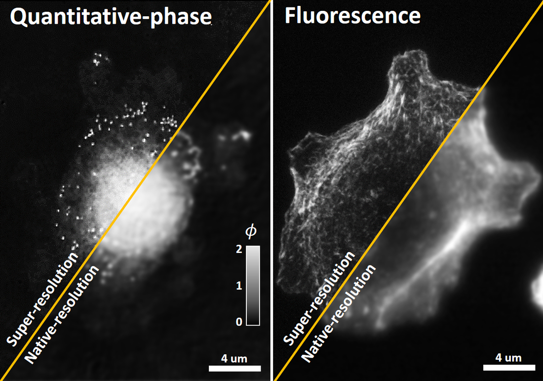
My previous work up till now had focused almost exclusively on projects targeting coherent super-resolution, with specific demonstrations targeting super-resolution of quantitative-phase (QP) and coherent scattering. These types of imaging systems are great for visualizing samples with endogenous contrast, which has applications in studies requiring for biophysical/biochemical analysis. However, for studies requiring visualization with molecular-specific contrast, fluorescence imaging reigns supreme. For this project, I wanted to develop an optical system that enabled efficient multimodal super-resolution with both fluorescence and QP. The hope was that this would make super-resolution more accessible to biologists who do not necessarily use either fluorescence or QP
question for this project: Can we develop an imaging system to allow multimodal quantitative-phase and fluorescent super-resolution?
question for this project: Can we develop an imaging system to allow multimodal quantitative-phase and fluorescent super-resolution?
Methods...
Because SIM has now been demonstrated for coherent super-resolution (based on my previous work), the obvious question is whether an imaging system can be outfitted with a SIM module to enable multimodal super-resolution. Furthermore, I wanted to extend this multimodal super-resolution capability into 3D. The hope is that biologists would be excited about this new system that can efficiently visualize multimodal fluorescent and QP 3D volumes at super-resolution scales - this could enable all sorts of intracellular studies to be done cohesively and thoroughly.
I built a new microscope system which used two 1.4 NA Zeiss objectives configured in transmission mode, as shown in Fig. 1 below with a pretty exhaustive description, and used a dichroic mirror to separate the coherent scatter from the fluorescent emission. Furthermore, I finally made a few additional key hardware changes, such as using a spatial-light-modulator (SLM) to allow automated structured pattern generation as well as a automated piezo-stage for automated z-scanning! Yay for automating image collection!
3D multimodal super-resolution system
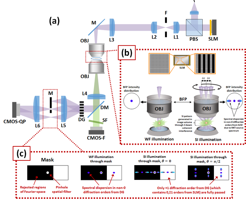
(a) Optical system consolidates conventional fluorescent and coherent structured illumination (SI) microscopies to enable sub-diffraction resolution quantitative-phase (QP) and fluorescent imaging. (b) Conventional widefield illumination is achieved when all the SLM pixels are turned ‘ON’. Sinusoidal structured illumination is achieved when the SLM is programmed to display a sinusoidal pattern. (c) The diffracted excitation beam from the sample is spectrally separated from the fluorescence, and is fed through a diffraction-phase-microscope (DPM). An asymmetric mask (M) is used to physically completely block the −1st diffraction order from DG while passing the + 1st diffraction order.
Results
The results from this project looked pretty good - it was my first time using a high-NA microscope objective, and it made a world of difference! Though this particular microscope was not built for speed (it was my first time foraying into automated machinery), there is no reason that an optical system with significantly faster hardware could not also be built for high-speed real-time imaging. But for now, I used this current system to image fixed biological cell samples.
The particular cells I imaged (A549) were fluorescently tagged for F-actin. With 3D fluorescent SIM, the F-actin filaments clearly stand out - however, no information about the other components are visible (as expected, since they were not fluorescently tagged). However, with super-resolved QP imaging, you can clearly see the nuclear region, high-density vesicles, and general cytoplasm and cell-body. In fact, you can register the different information channels on each other to calculate the 3D positions of all the components. Pretty cool!
Below, we show an example of multimodal super-resolution imaging of A549 cells, which are from a human adenocarcinoma breast cancer cell line. The fluorescence image shows the F-actin components within the cell while the QP image shows the general cell morphology, from which the nucleus, vesicles, and cell boundary are clearly visible and can be outlined. Thus, after some easy registration to account for inter-camera differences, we can figure out the relative positions of the intracellular components to each other, regardless of modality, as shown below by the green, blue, red outlines for the cellular periphery, vesicle clusters, and nucleus, respectively.
Of course, because all this is super-resolved in 3D, we also show SI vs widefield (WF) fluorescent/QP signal at different layers through the cell. In the case of the QP image volume, we see that the visibility of the vesicles quickly diminish with increasing depth only in the SI-volume, not in the conventional WF-QP image. In the fluorescent image, we can see the F-actin filaments clearly undergoing morphological change with increasing depth, while the conventional WF fluorescent image just shows a hazy signal at all depths. In both cases, we see that SI enables 3D optical sectioning, and thus allows 3D multimodal super-resolution!
Multimodal visualization of A549 cells with fluorescent/QP super-resolution
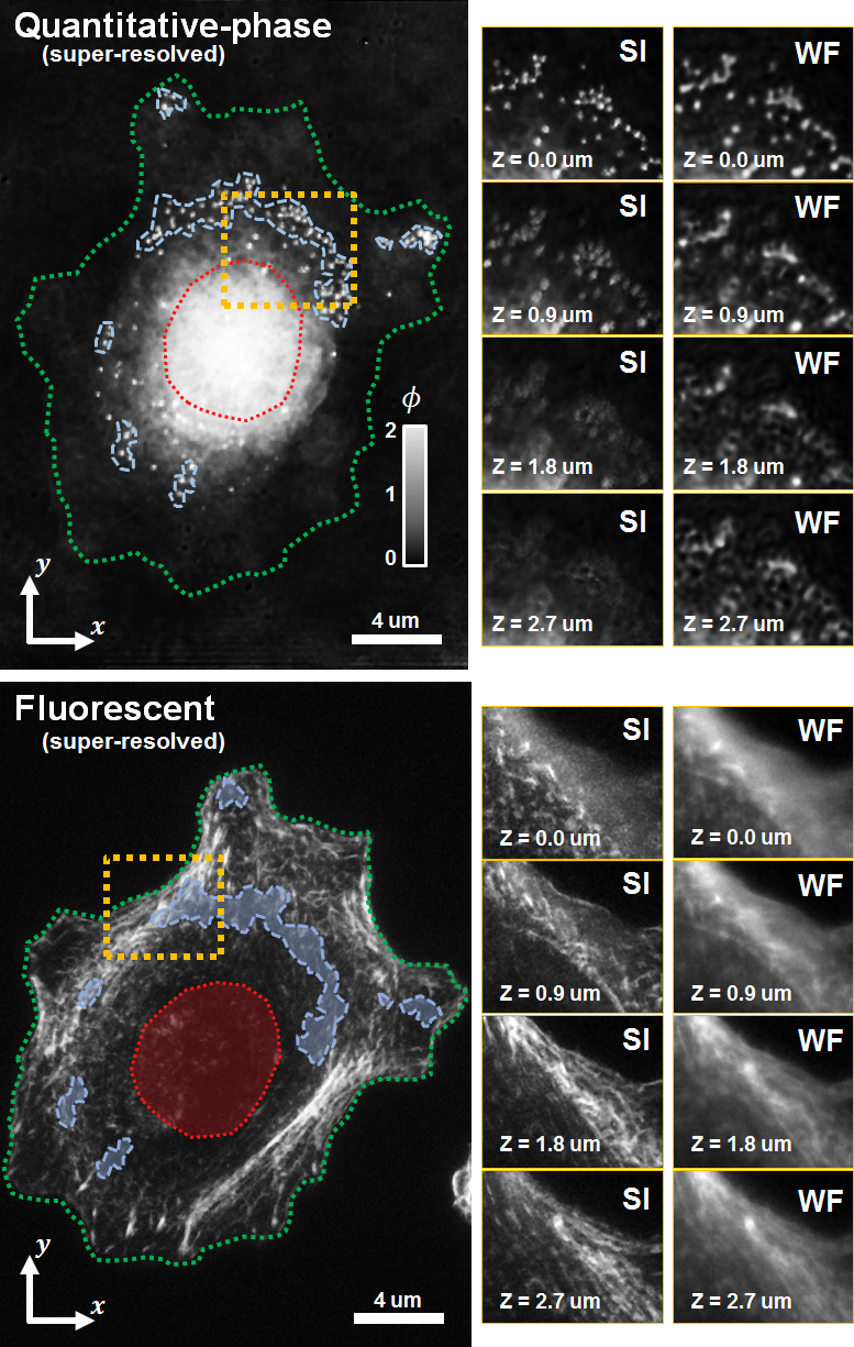
3D visualization capabilities of QP and fluorescent imaging are compared when visualizing an individual A549 cell. SI-enhanced QP sub-diffraction resolution is clearly evident from a zoom of the region outlined in yellow in which clearly resolves vesicles with SI- but not WF- QP imaging. 3D imaging capabilities between SI and WF are demonstrated by observing that the sharp QP signal from the vesicles hare attenuated with increasing defocus, indicating optical depth sectioning. In contrast, the WF volume showed the QP signal from the high phase-delay structures diffracting out into the defocused planes, leading to diffraction artifacts indistinguishable from in-focus QP signal. Fluorescent resolution was also enhanced when comparing SI to WF imaging, respectively. Defocused planes show that SI fluorescence imaging demonstrates clear optical sectioning and shows the actin morphology undergoing clear organizational changes through different depths of the cell. In contrast, defocused planes through the WF fluorescence volume show a strong defocused signal throughout the volume stack, which hinders visualization of important high resolution features.
Project publications