Structured illumination for super-resolution quantitative-phase microscopy
Quantitative-phase microscopy (QPM) via holography is a great way to visualize transparent, unlabelled samples that would otherwise be almost invisible with conventional brightfield microscopy. However, holography is still subject to a diffraction limit just as any other imaging system. I wanted to see if the QPM resolution achieved via holography could also be improved via structured illumination in the same way Gustaffson initially proposed in the early 2000s (though he limited his works to fluorescence imaging). In contrast to my previous project, where nonlinearities arose from coherent, intensity-based imaging, holography allows direct electric-field detection, which is a linear coherent imaging process. Hence, Gustaffson's super-resolution framework should directly allow for QPM with holographic detection!.
question for this project: Can structured illumination be used for super-resolution of quantiative-phase imaging
question for this project: Can structured illumination be used for super-resolution of quantiative-phase imaging
Methods...
Off-axis holography is well-associated with a loss in resolution due to unused k-space pixels. In typical applications where the user simply wants QP imaging capabilities, these unused k-space pixels are usually considered to simply be an unfortunate disadvantage of the system. However, if the desired outcome is to enable simultaneous, single-shot multimodal imaging, these unused k-space pixels are prime candidates with which to multiplex the information content from other modalities. The trick is to find an optical way to do so!
Off-axis holography is well-associated with a loss in resolution due to unused k-space pixels. In typical applications where the user simply wants QP imaging capabilities, these unused k-space pixels are usually considered to simply be an unfortunate disadvantage of the system. However, if the desired outcome is to enable simultaneous, single-shot multimodal imaging, these unused k-space pixels are prime candidates with which to multiplex the information content from other modalities. The trick is to find an optical way to do so!
Results
The results from this project were pretty promising! As mentioned above, the trick was to find a way to multiplex the information from the fluorescent and QP modalities into different regions of k-space. To do this, the key breakthrough came in the realization that this could be possible if each modality was independently modulated by a carrier wave, pre-set to a unique configuration, before being imaged onto the camera. This concept is actually quite ubiquitous in our everyday world, in the form of AM modulation in typical radio! So how do we modulate the different modalities? Gratings are the solution! The natural periodicity of the gratings will work well to modulate the multimodal info into different regions of k-space!
Below, I show the developed imaging system that uses ronchi gratings to do exactly this. A few things to note: (1) each modality is spectrally (color) separated from each other - this allows easy spectral separation between the different modalities, which is necessary for modulation, before being rejoined all together on the camera. (2) Each ronchi grating occupies a unique rotation angle - this each modality content to be modulated to a unique spot in k-space to minimize overlap. (3) Multimodal overlap needs to be minimized for easy separation and subsequent retrieval! I show the preferred k-space modulation locations that I chose for multiplexing 1 QP channel with 2 fluorescent channels. I also show a simultaneously acquired multimodal data-set of a COS-7 cell with QP and fluorescent (nuclear and F-actin staining) contrast.
|
Optical system for multiplexed imaging
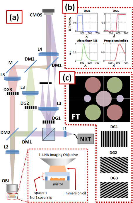
(a) Optical schematic of the multiplexed imaging system, (b) Spectra for the dichroic mirrors and fluorescent stains used in the system (c) Example of Fourier separation between the system's different modalities using the rotated gratings DG1, DG2, and DG3.
|
Multiplexed visualization of COS-7 cells with fluorescent/QP
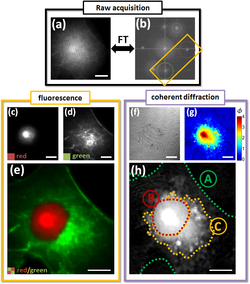
(a) Raw acquisition and (b) associated Fourier spectrum. Digital filters used for fluorescence and QP retrieval are outlined. Fluorescence reconstructions are shown from the (c) red and (d) green color channels, showing nucleus and F-actin, respectively. (f) Amplitude and (g) QP image reconstructions are also shown. (h) Gray-scale QP image with labelled cell body, nucleus, and endoplasmic reticulum is shown. Scale bar is 10 um.
|
Project publications
